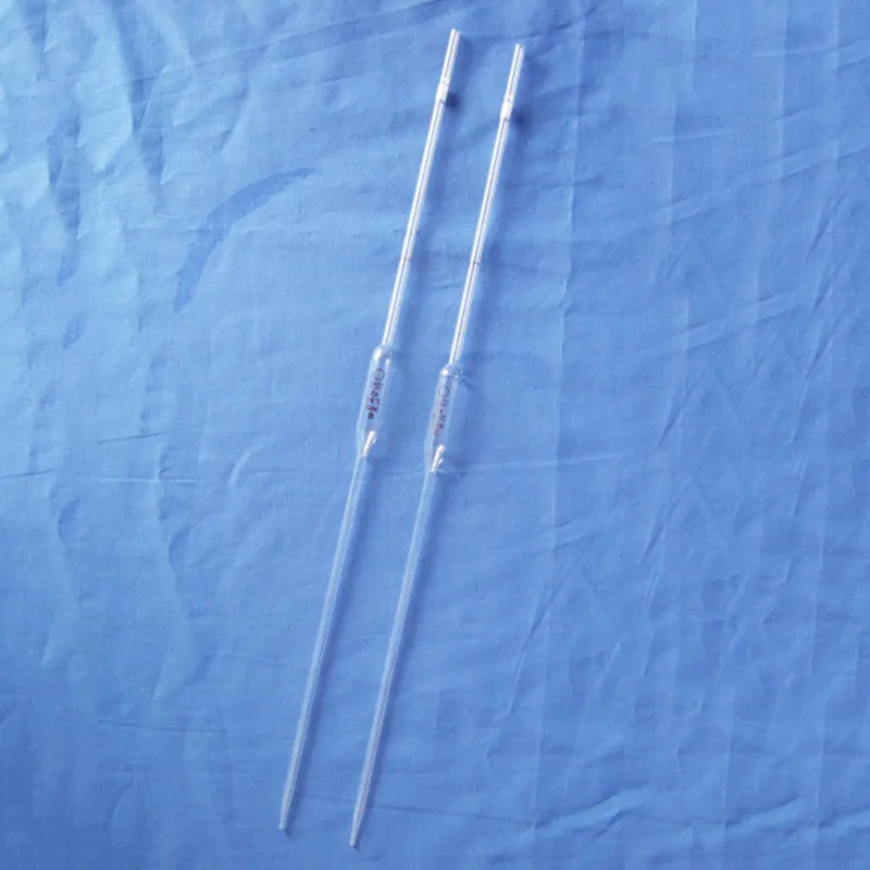
With multi-layer coated optics, the ear microscope delivers better light transmission and image contrast. Ergonomic design allows for comfortable long-term use. The smooth stage movement and fine focusing system provide sensitive slide control for accurate analysis. The ear microscope can be used with image capture systems for recording and sharing information, supporting both live observation and digital research workflows in the classroom and lab.

Across the worlds of science, industry, and education, the ear microscope enables research at the microscopic level. It is an essential tool in medical diagnosis to analyze blood, tissues, and pathogens. Environmental scientists apply the ear microscope to determine bacteria and microalgae that indicate water levels of quality. In materials science, it enables nanostructure analysis and the identification of defects. Art conservators apply the ear microscope to analyze pigments and varnish layers. Its ability to produce accurate, detailed imagery makes it a valuable resource in continuing discovery and research development.

The ear microscope of the future will be to expand its analytical power. Future models will integrate optical accuracy with the enhancement of the computer, creating hybrid devices with real-time analysis functions. Automation will ease routine operations, making laboratory workflow more efficient. The ear microscope will also be able to integrate cloud-based platforms for real-time sharing of data and remote access. Environment-friendly technology development will yield models that are energy-efficient without sacrificing precision but reduce environmental impact.

To continue functioning optimally, the ear microscope must be treated to regular maintenance with attention to detail. Clean lenses with soft strokes using microfiber cloths or dedicated wipes. Avoid spraying cleaners directly on the optics. Keep the stage and focus assembly residue and corrosion free. Always shut down when cleaning electrical components. When storing, cover the ear microscope and place it in a dry, temperature-controlled environment. Periodic service inspections will ensure accurate focusing, smooth operation, and long-term durability.
A ear microscope is able to closely study microorganisms, tissue, and materials and is thus a fundamental instrument in laboratories and classrooms. It operates by bending light or electron rays to enlarge specimens to appear gigantic many times magnification. The ear microscope has been enhanced with developments in optics to enable brighter, clearer, and digital-imaging-assisted magnification. In academic research work as well as industrial inspection, a ear microscope enables accurate analysis, recording, and examination of complex microscopic realms.
Q: What is the lifespan of a microscope? A: With proper care and maintenance, a microscope can last for many years, providing consistent optical performance and stability. Q: How does the objective lens affect image quality in a microscope? A: The objective lens determines magnification and resolution; high-quality lenses produce sharper, more accurate images of specimens. Q: Can a microscope be used to view live specimens? A: Yes, many microscope models support live-cell observation, allowing users to study biological processes in real time under controlled conditions. Q: What is the function of the condenser in a microscope? A: The condenser focuses light onto the specimen, enhancing illumination and improving contrast for clear image viewing. Q: How should a microscope be transported safely? A: Carry the microscope with both hands—one under the base and one on the arm—to prevent damage or misalignment of delicate parts.
The microscope delivers incredibly sharp images and precise focusing. It’s perfect for both professional lab work and educational use.
This ultrasound scanner has truly improved our workflow. The image resolution and portability make it a great addition to our clinic.
To protect the privacy of our buyers, only public service email domains like Gmail, Yahoo, and MSN will be displayed. Additionally, only a limited portion of the inquiry content will be shown.
We’re interested in your delivery bed for our maternity department. Please send detailed specifica...
Hello, I’m interested in your centrifuge models for laboratory use. Could you please send me more ...
E-mail: [email protected]
Tel: +86-731-84176622
+86-731-84136655
Address: Rm.1507,Xinsancheng Plaza. No.58, Renmin Road(E),Changsha,Hunan,China