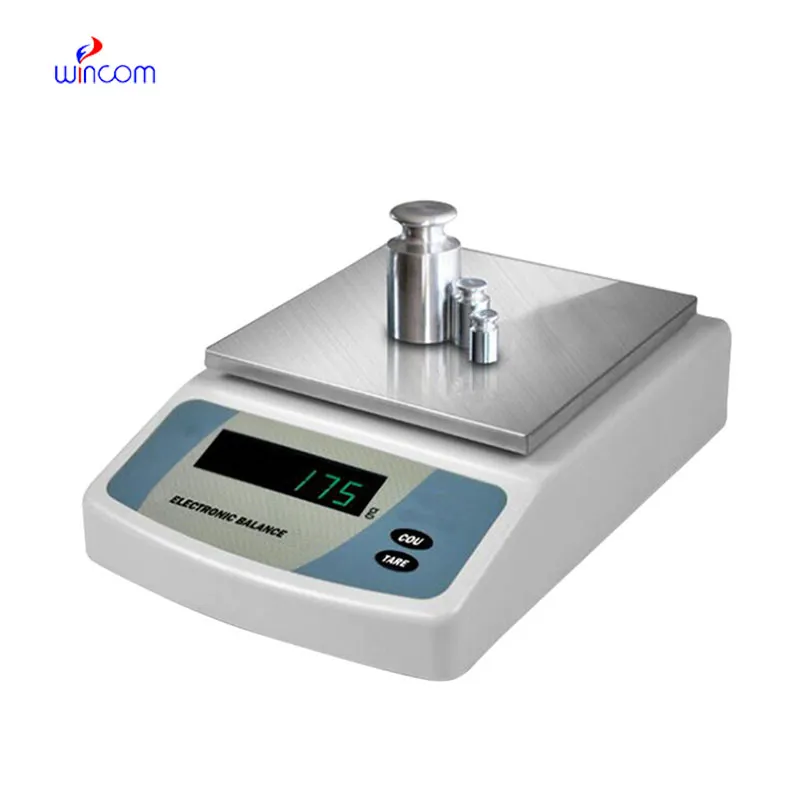
In the laboratories of hospitals and clinics, electronic balancing is responsible for the precise weighing of patient samples, reagents, and pharmaceutical ingredients. Being highly precise, it minimized sample preparation errors and that is a good support for analytical results that can be reproduced. Laboratory techs apply electronic balancing in the processes of quality control, method validation, and even daily operations. Reliable diagnostics, efficient laboratory workflows, and high-quality research and medical testing are the consequences of the accuracy and consistency maintained by electronic balancing.

In pathology laboratories, electronic balancing finds its usage during the staining compounds preparation and the tissue processing additives application. The proper mass measurement guarantees the same composition of the reagent, and this, in turn, affects the performance of the stain and the interpretation under the microscope. This application helps to maintain the same standards in pathology workflows and minimizes the differences between test batches. By using strict preparation conditions, electronic balancing plays a role in the stability of diagnosis in hospital pathology departments.
The future development of electronic balancing for laboratory and hospital use will increasingly depend on sustainability. Operational consumption will be lowered by using energy-efficient parts and incorporating optimized power management. The progression of these technologies will not only make analytical devices compatible with hospital sustainability but also ensure dependable performance in nonstop laboratory operations.

electronic balancing in clinics is, however, maintained through regular performance verification conducted in the laboratories. Certified test weights are used to prove the reliability of the measurements with the passage of time. Verification activities being documented also implies good traceability and makes it easier for internal audits to take place. By making verification part of the routine maintenance, hospitals make sure that electronic balancing still gives trustworthy results for research and diagnostic workflows.
To formulate a drug, electronic balancing is used by the pharmaceutical laboratories to weigh active ingredients and excipients. Precise and reliable measurements not only guarantee the correctness of the dosage but also satisfy the requirements of the regulators. The laboratory personnel use electronic balancing for quality control, batch verification, and stability testing. Its accuracy aids in the production of medicines that are reliable, thus minimizing errors in the production. Adoption of electronic balancing into workflow has helped pharmaceutical labs not only to keep their quality standards high but also to make sure that patients are safe by providing the exact analytical measurements.
Q: What maintenance does an Analytical Balance require? A: A periodic cleaning, checking of the calibration, and also verifying the performance are all necessary. Q: Can an Analytical Balance handle continuous daily use? A: Yes, provided that the correct laboratory conditions and rules are followed. Q: Why is leveling important for an Analytical Balance? A: The accuracy and repeatability of the measurements depend on proper leveling. Q: Can Analytical Balances be connected to laboratory systems? A: Most of the models allow connectivity with laboratory information systems. Q: Are Analytical Balances sensitive to vibration? A: Yes, stable weight readings can be disturbed by vibrations.
The water bath performs consistently and maintains a stable temperature even during long experiments. It’s reliable and easy to operate.
I’ve used several microscopes before, but this one stands out for its sturdy design and smooth magnification control.
To protect the privacy of our buyers, only public service email domains like Gmail, Yahoo, and MSN will be displayed. Additionally, only a limited portion of the inquiry content will be shown.
We are planning to upgrade our imaging department and would like more information on your mri machin...
We’re looking for a reliable centrifuge for clinical testing. Can you share the technical specific...
E-mail: [email protected]
Tel: +86-731-84176622
+86-731-84136655
Address: Rm.1507,Xinsancheng Plaza. No.58, Renmin Road(E),Changsha,Hunan,China

 af
af
 es
es
 ar
ar
 tr
tr
 sw
sw
 pt
pt
 th
th
 ur
ur
 bn
bn
 ne
ne
 vi
vi
 km
km
 lo
lo
 de
de
 ru
ru
 fi
fi
 nl
nl
 fa
fa
 fr
fr
 ko
ko