The fetal doppler hand held, being designed for flexibility, brings the user a variety of imaging modes such as B-mode, M-mode, and color Doppler to use. Because of its small size, it can be easily moved from one place to another and that is why it can be used for examining patients in bed. The fetal doppler hand held provides good imaging quality, which can be depended upon in cases of routine diagnostics, fieldwork, and emergency medical interventions.

The fetal doppler hand held has demonstrated its irreplaceable nature in prenatal screening, cardiovascular diagnostics, and overall health evaluations.The fetal doppler hand held is a technique that evaluates organ function, reveals pathological changes, and supports medical education by providing live imaging demonstrations. The fetal doppler hand held technology gives doctors the ability to perform accurate and instantaneous assessments in a variety of clinical situations.

The future of the fetal doppler hand held may include enhancements based on artificial intelligence algorithms that can improve images and offer automatic measurements. Better transducer technologies may offer higher resolution and increased sensitivity. The fetal doppler hand held may have an increasing role to play in personalized medicine because it may offer continuous monitoring capabilities.

The fetal doppler hand held require a certain set of procedures when it comes to handling them. The images on the display should always be cleaned with soft, lint-free cloths. The probe membranes should always be checked for the absence of cracks. The fetal doppler hand held also require calibration verification.
The fetal doppler hand held is an essential diagnostic modality in the modern healthcare system, permitting the non-invasive imaging of internal organs and tissues. By transmitting sound waves and reading their echoes, it provides real-time data on physiology. The fetal doppler hand held makes precise diagnoses feasible in all specialties, improving clinical decision-making and patient confidence.
Q: What makes the ultrasound scannert effective for diagnostic imaging? A: Its high-frequency sound wave technology allows accurate visualization of internal body structures in real time. Q: How portable is the ultrasound scannert? A: The device features a compact and lightweight design, allowing easy movement between clinical departments. Q: What types of probes are compatible with the ultrasound scannert? A: It supports multiple probe types, including linear, convex, and phased array probes for varied diagnostic needs. Q: Does the ultrasound scannert require special training to operate? A: Basic technical training is recommended to maximize its imaging performance and functionality. Q: How long can the ultrasound scannert operate continuously? A: It is designed for extended use with efficient cooling systems and stable power performance.
We’ve used this centrifuge for several months now, and it has performed consistently well. The speed control and balance are excellent.
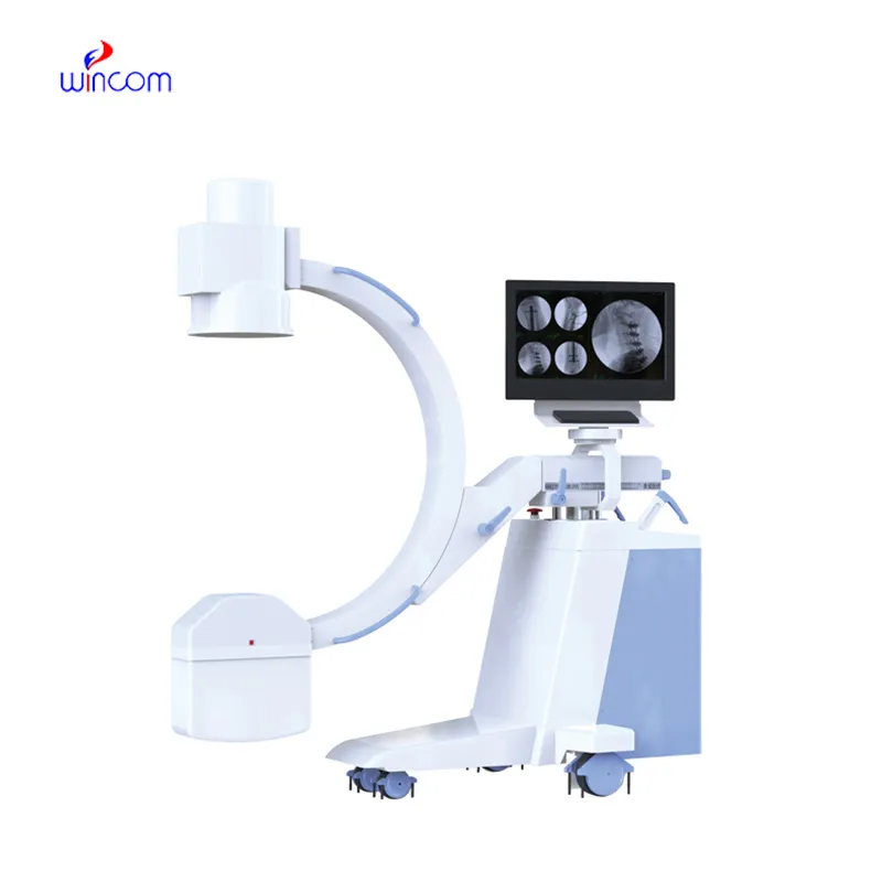
This x-ray machine is reliable and easy to operate. Our technicians appreciate how quickly it processes scans, saving valuable time during busy patient hours.
To protect the privacy of our buyers, only public service email domains like Gmail, Yahoo, and MSN will be displayed. Additionally, only a limited portion of the inquiry content will be shown.
Hello, I’m interested in your centrifuge models for laboratory use. Could you please send me more ...
We are planning to upgrade our imaging department and would like more information on your mri machin...
E-mail: [email protected]
Tel: +86-731-84176622
+86-731-84136655
Address: Rm.1507,Xinsancheng Plaza. No.58, Renmin Road(E),Changsha,Hunan,China

 af
af
 es
es
 ar
ar
 tr
tr
 sw
sw
 pt
pt
 th
th
 ur
ur
 bn
bn
 ne
ne
 vi
vi
 km
km
 lo
lo
 de
de
 ru
ru
 fi
fi
 nl
nl
 fa
fa
 fr
fr
 ko
ko