The fetal doppler machine has been designed considering the needs of modern health care, providing uninterrupted performance with its rapid image acquisition and high-definition visualization. The robust outer casing and sophisticated temperature control system will make sure that the device continues to work and be trustworthy. Also, the fetal doppler machine is a device that aids the long-term data archiving process for efficient medical record management.

The fetal doppler machine has become a necessity for internal medicine as it provides real-time imaging during fluid drainage and biopsy guidance. It finds a place in critical care too for instant bedside evaluations. The fetal doppler machine is also utilized by veterinary surgeons to monitor the health of a patient animal, thus proving its utility beyond human medicine.

Through continued innovations in digital technology, the fetal doppler machine can be expected to improve and extend its applications within preventive medicine and telemedicine. The next generation of such technologies will facilitate collaboration among experts in real-time using cloud-imaging solutions. The fetal doppler machine can also work within wearables that include biosensors.

Proper upkeep of the fetal doppler machine helps maintain both the safety of the users as well as the durability of the equipment. The equipment's ventilation and power components must also be regularly inspected for evidence of obstruction and wear. In order to maintain the continued high-quality output of images from the fetal doppler machine, it must be properly maintained.
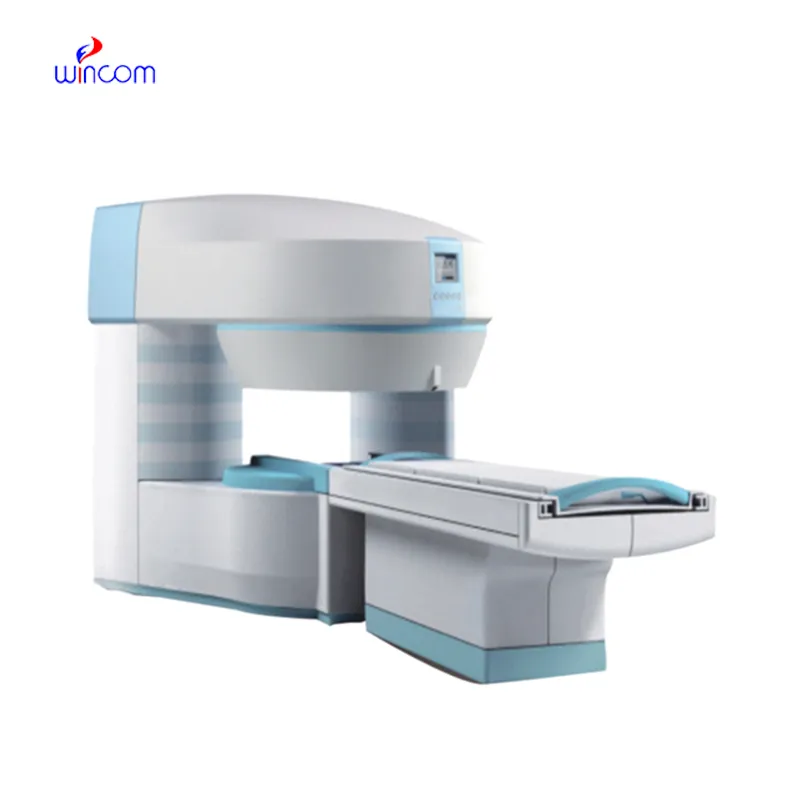
The fetal doppler machine represents an advanced form of medical imaging technology that transforms sound waves into high-definition visual data. It is widely used for evaluating organ health, tracking fetal development, and detecting vascular conditions. The fetal doppler machine ensures real-time monitoring and fast diagnostic results, supporting effective clinical workflows.
Q: What are the main maintenance requirements for the ultrasound scannert? A: Regular cleaning, proper probe handling, and scheduled inspections help maintain optimal performance. Q: How often should the ultrasound scannert be calibrated? A: Calibration frequency depends on usage levels, but periodic professional checks are recommended. Q: Is the ultrasound scannert suitable for pediatric use? A: Yes, it provides gentle, non-invasive imaging ideal for neonatal and pediatric diagnostics. Q: Does the ultrasound scannert support wireless connectivity? A: Many models include Wi-Fi or Bluetooth features for data sharing and device integration. Q: What materials are used in the ultrasound scannert construction? A: It is built with durable medical-grade components designed to withstand continuous clinical use.
The microscope delivers incredibly sharp images and precise focusing. It’s perfect for both professional lab work and educational use.
The centrifuge operates quietly and efficiently. It’s compact but surprisingly powerful, making it perfect for daily lab use.
To protect the privacy of our buyers, only public service email domains like Gmail, Yahoo, and MSN will be displayed. Additionally, only a limited portion of the inquiry content will be shown.
Hello, I’m interested in your water bath for laboratory applications. Can you confirm the temperat...
I’d like to inquire about your x-ray machine models. Could you provide the technical datasheet, wa...
E-mail: [email protected]
Tel: +86-731-84176622
+86-731-84136655
Address: Rm.1507,Xinsancheng Plaza. No.58, Renmin Road(E),Changsha,Hunan,China

 af
af
 es
es
 ar
ar
 tr
tr
 sw
sw
 pt
pt
 th
th
 ur
ur
 bn
bn
 ne
ne
 vi
vi
 km
km
 lo
lo
 de
de
 ru
ru
 fi
fi
 nl
nl
 fa
fa
 fr
fr
 ko
ko