Hospitals and biomed research centers employ gas liquid chromatography mcat that help optimize patient testing and lab work. By being able to distinguish, measure, and analyze drugs, metabolites, and biomolecules, gas liquid chromatography mcat is a necessary tool in patient testing. Lab professionals incorporate gas liquid chromatography mcat into lab work on a daily basis. Reproducibility and analytical ability make gas liquid chromatography mcat an irreplaceable tool in assisting with patient testing.

Hospital laboratories depend on gas liquid chromatography mcat for identifying minute quantities of pharmaceuticals and therapeutic agents in difficult-to-analyze biological samples. Its use spans drug compliance testing, pharmacokinetics profiling, and tracking medications after surgery. The laboratory personnel can rely on it for exact measurement, thus increasing the efficiency of clinical treatment.
gas liquid chromatography mcat is assigned to become an important player in translational research which is being conducted in hospitals. Among the future developments are the combined detection systems, quicker analysis cycles, and improved reproducibility. gas liquid chromatography mcat will be the mainstay of hospitals' molecular profiling and drug testing along with patient monitoring thus facilitating hospital diagnostics and personalized medicine research.
gas liquid chromatography mcat will require regular maintenance to be kept up in order to continue providing precise measurements in medical laboratories. After every use, the technicians should flush the columns, check the seals, and inspect the tubing for wear and tear and ensure that the detector is working. Regular calibration and good solvent management decrease the chances of system damage and increase the consistency of the results. Good care and maintenance not only increase the efficiency of the laboratory but also help in providing reliable diagnostics and maintaining the instruments for hospital applications.
gas liquid chromatography mcat is employed by laboratories in hospitals and research centers to keep control over their analytical quality in a manner that is non-stop. It works by separations of different chemicals in complex mixtures, pinpointing the impurities, and very accurately quantifying the concentrations. Technicians in the laboratory depend on gas liquid chromatography mcat for the purposes of method verification, calibration, and validation of techniques for analysis. It is in clinical and pharmaceutical labs that the instrument changes the generated data into accurate and reproducible forms. Its high-resolution separation capacity is utilized by both modern testing and up-to-date research projects. gas liquid chromatography mcat is given the credit of being the backbone instrument in laboratory operations by providing detailed results that are consistent, thus being the source of reliable analysis and supporting the whole medical and experimental research by maintaining its integrity.
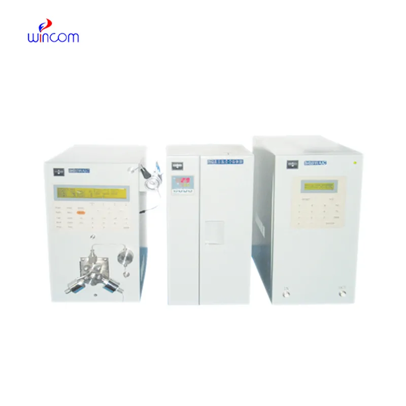
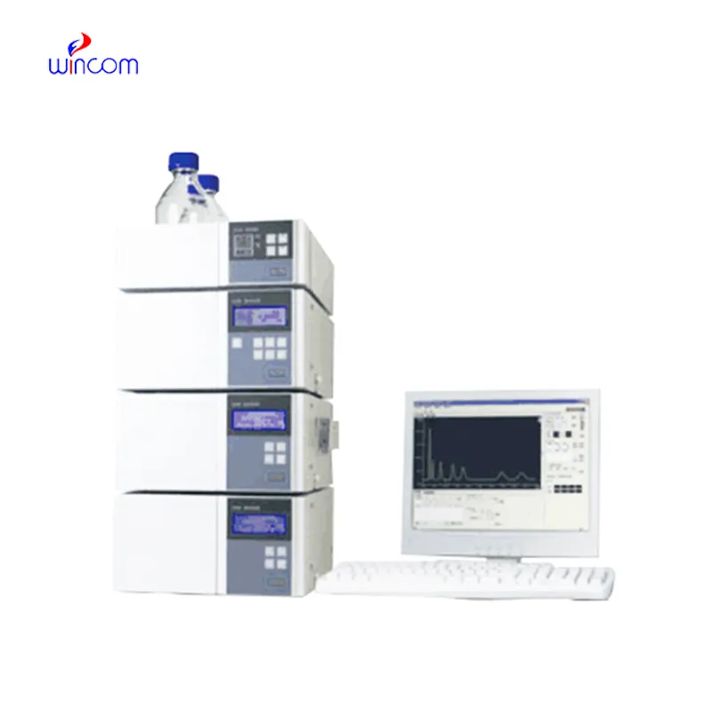
Q: Do you need special training for HPLC operation? A: The answer is yes, training is a prerequisite to accurately and safely using pumps, columns, and detectors. Q: What type of maintenance does HPLC have? A: It requires cleaning, flushing, and inspection of all components as well as calibrating. Q: Is it possible to use HPLC in drug monitoring? A: Sure, it is a common practice in hospitals to monitor the levels of therapeutic drugs and also to identify metabolites in the samples taken from the patients. Q: What is the duration of analysis using HPLC in a typical case? A: The analysis time can range from a few minutes to more than an hour depending on the nature of the sample and the kind of column used. Q: Is HPLC a good choice for environmental testing? A: Yes, it can be used to find out the presence of pollutants, pesticides, and other harmful substances in water, soil, and air samples.
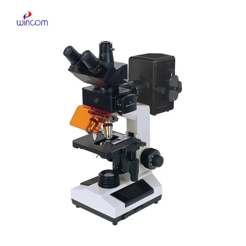
I’ve used several microscopes before, but this one stands out for its sturdy design and smooth magnification control.
We’ve been using this mri machine for several months, and the image clarity is excellent. It’s reliable and easy for our team to operate.
To protect the privacy of our buyers, only public service email domains like Gmail, Yahoo, and MSN will be displayed. Additionally, only a limited portion of the inquiry content will be shown.
We’re currently sourcing an ultrasound scanner for hospital use. Please send product specification...
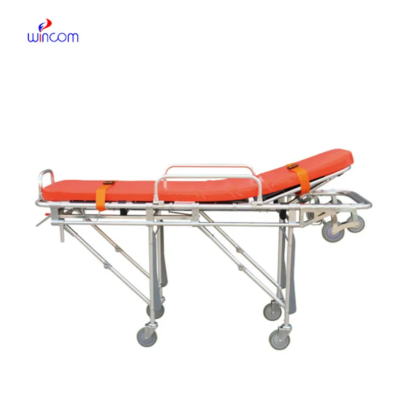
Could you share the specifications and price for your hospital bed models? We’re looking for adjus...
E-mail: [email protected]
Tel: +86-731-84176622
+86-731-84136655
Address: Rm.1507,Xinsancheng Plaza. No.58, Renmin Road(E),Changsha,Hunan,China

 af
af
 es
es
 ar
ar
 tr
tr
 sw
sw
 pt
pt
 th
th
 ur
ur
 bn
bn
 ne
ne
 vi
vi
 km
km
 lo
lo
 de
de
 ru
ru
 fi
fi
 nl
nl
 fa
fa
 fr
fr
 ko
ko