hplc analyse enables labs to separate and analyze intricate mixtures with utmost precision. Through a seamless connection with current detectors, the method provides detailed profiling of both chemical and biological substances. The researchers and therapists trust hplc analyse for the purposes of monitoring outcomes of experiments, method development, and cross-analyses accuracy. Its strength in dealing with various kinds of samples renders it an indispensable device in both the research and the clinical settings, thus improving reproducibility and backing up the struggling with more complex scientific and medical inquiries.

hplc analyse finds extensive application in hospital laboratories for monitoring drugs therapeutically. It provides precise determination of drug levels in patients' samples, thus making safe and effective dosing possible. Metabolites are tracked, treatment progress is assessed, and unexpected drug interactions are detected by the laboratory personnel. Its high accuracy and reproducibility facilitate both medical decision-making and research, hence, hplc analyse becomes an indispensable instrument in taking care of patients and analyzing the medical field.

In hplc analyse, the evolution is probably going to be through miniaturization and portability hplc analyse is the main feature of the future hospital and laboratory. These advancements will let bedside or point-of-care analysis, thus, improving hospital diagnostics and reducing turnaround times. The future highlights quickness, highly reproducible measurements, and still good accuracy in patient monitoring and laboratory research.

Systematic attention on the system components is necessary for the running of hplc analyse in hospital and research labs. To prevent contamination and pressure problems, flushing of columns, seal replacements, and tubing inspections should be done regularly. Regular calibration of detectors and documentation of maintenance procedures should be done by laboratory technicians. The instruments' life is prolonged by consistent care and monitoring, which also lead to accurate sample analysis and support the reliability of laboratory operations both for clinical and experimental purposes.
hplc analyse is a standard method in diagnostic laboratories of hospitals to keep an eye on patients’ biochemical and therapeutic figures. It quantifies drugs, hormones, and small molecules accurately. hplc analyse speeds up the clinical decision-making processes of physicians and facilitates treatment modifications by supplying them with quick and precise results. It is used by hospital labs for basic patient testing, pharmacokinetic studies, and special analyses. The method’s high reproducibility makes certain that the outcomes are consistent, whereas its versatility allows for the support of many clinical applications. hplc analyse has turned into an irreplaceable instrument in hospital diagnostics, which not only enhances patient management but also provides healthcare professionals with thorough molecular information.
Q: What are the main parts of a microscope? A: The key components include the eyepiece, objective lenses, stage, focusing knobs, and illumination system, all working together to magnify and clarify specimens. Q: How do you clean the lenses of a microscope? A: Lenses should be cleaned using soft lens paper or microfiber cloth with a small amount of lens cleaner to avoid scratching or damaging optical coatings. Q: What magnification levels can a microscope achieve? A: Depending on the model, a microscope can typically achieve magnifications ranging from 40x to over 1000x for detailed observation of microscopic structures. Q: Why is light adjustment important in a microscope? A: Proper light adjustment ensures accurate contrast and brightness, allowing clear observation without distortion or glare during viewing. Q: Can a microscope be used for educational purposes? A: Yes, microscopes are widely used in classrooms and laboratories to teach students about biology, materials science, and microscopic analysis.
The delivery bed is well-designed and reliable. Our staff finds it simple to operate, and patients feel comfortable using it.
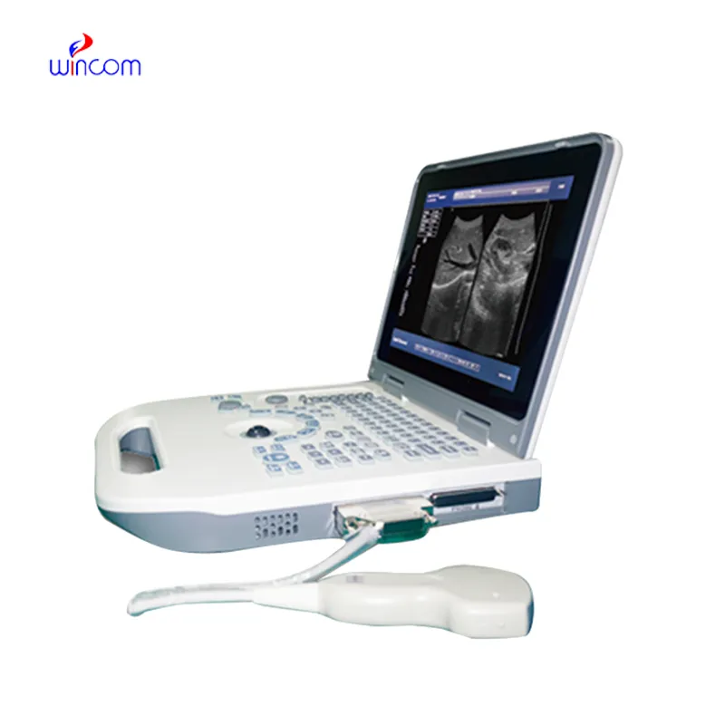
This ultrasound scanner has truly improved our workflow. The image resolution and portability make it a great addition to our clinic.
To protect the privacy of our buyers, only public service email domains like Gmail, Yahoo, and MSN will be displayed. Additionally, only a limited portion of the inquiry content will be shown.
Hello, I’m interested in your centrifuge models for laboratory use. Could you please send me more ...
I’m looking to purchase several microscopes for a research lab. Please let me know the price list ...
E-mail: [email protected]
Tel: +86-731-84176622
+86-731-84136655
Address: Rm.1507,Xinsancheng Plaza. No.58, Renmin Road(E),Changsha,Hunan,China

 af
af
 es
es
 ar
ar
 tr
tr
 sw
sw
 pt
pt
 th
th
 ur
ur
 bn
bn
 ne
ne
 vi
vi
 km
km
 lo
lo
 de
de
 ru
ru
 fi
fi
 nl
nl
 fa
fa
 fr
fr
 ko
ko