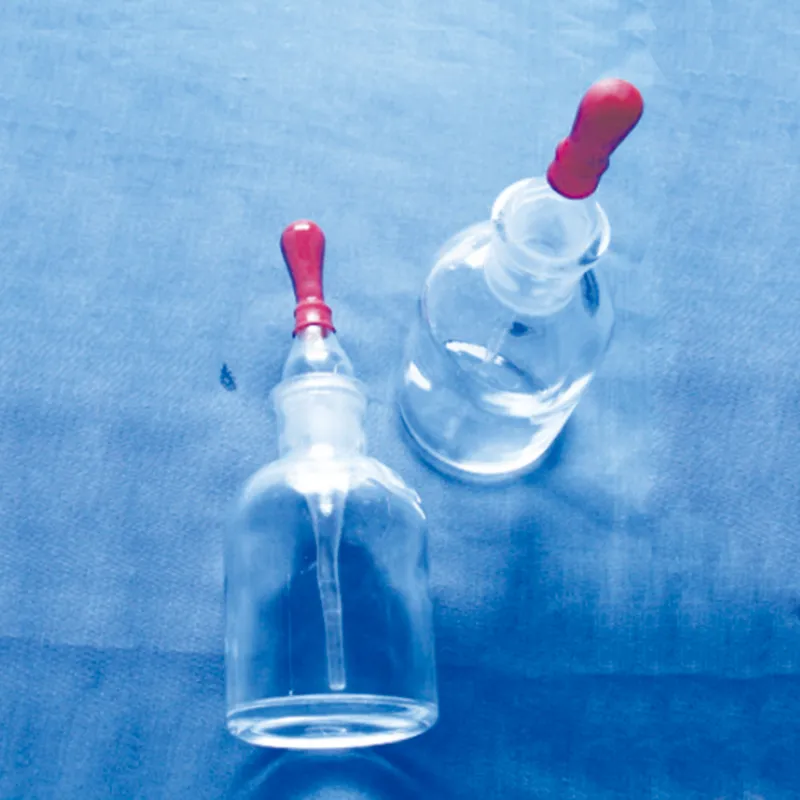
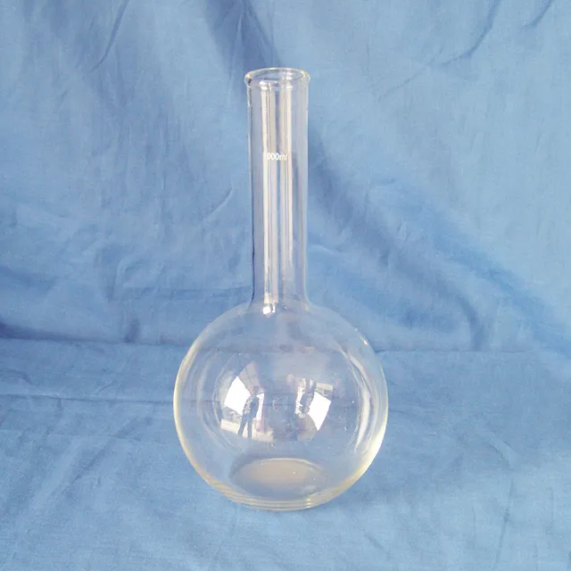
liquid chromatography description enables labs to separate and analyze intricate mixtures with utmost precision. Through a seamless connection with current detectors, the method provides detailed profiling of both chemical and biological substances. The researchers and therapists trust liquid chromatography description for the purposes of monitoring outcomes of experiments, method development, and cross-analyses accuracy. Its strength in dealing with various kinds of samples renders it an indispensable device in both the research and the clinical settings, thus improving reproducibility and backing up the struggling with more complex scientific and medical inquiries.

Hospital laboratories depend on liquid chromatography description for identifying minute quantities of pharmaceuticals and therapeutic agents in difficult-to-analyze biological samples. Its use spans drug compliance testing, pharmacokinetics profiling, and tracking medications after surgery. The laboratory personnel can rely on it for exact measurement, thus increasing the efficiency of clinical treatment.

In liquid chromatography description, the evolution is probably going to be through miniaturization and portability liquid chromatography description is the main feature of the future hospital and laboratory. These advancements will let bedside or point-of-care analysis, thus, improving hospital diagnostics and reducing turnaround times. The future highlights quickness, highly reproducible measurements, and still good accuracy in patient monitoring and laboratory research.

Proper handling and care of liquid chromatography description ensure continuous accuracy in the medical laboratory workflows. Cleaning of flow paths, checking detector response, and verifying pump performance are the essential maintenance tasks. Along with the column storage, solvent selection, and routine calibration, laboratory personnel must adhere to the manufacturer guidelines. Proper care enhances reproducibility, reduces downtime, and supports the consistent performance of the laboratory in hospitals and clinical research facilities.
liquid chromatography description is of utmost importance in biochemistry laboratories of both universities and hospitals. It makes detailed study of proteins, peptides, and metabolites possible through the separation of intricate mixtures. The application of it includes but is not limited to enzymatic analysis, biomarker detection, and data obtained through metabolomics. The sensitivity and reproducibility of the device guarantee genuine molecular profiles. Lab technicians make use of liquid chromatography description to conclude their experiments and provide evidence for scientific publications. Its accuracy and versatility give biochemistry labs the ability to perform cutting-edge research in molecular mechanisms, disease pathways, and therapy targets thus, it becomes an indispensable tool for both analytical and clinical lab investigations.
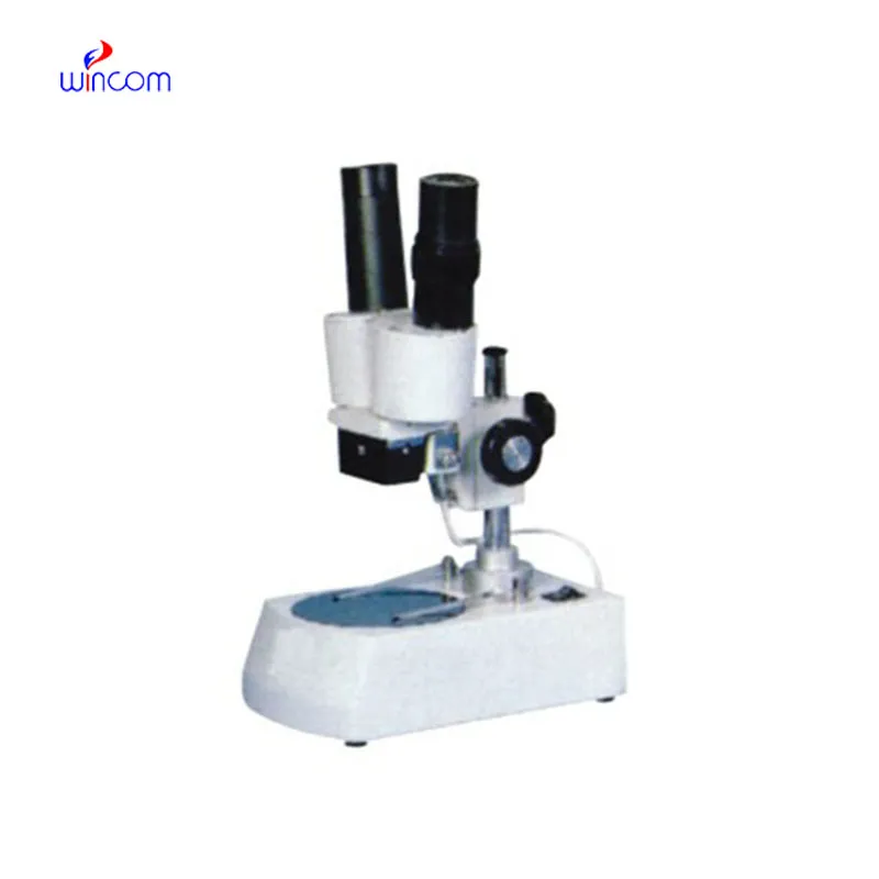
Q: What are the main parts of a microscope? A: The key components include the eyepiece, objective lenses, stage, focusing knobs, and illumination system, all working together to magnify and clarify specimens. Q: How do you clean the lenses of a microscope? A: Lenses should be cleaned using soft lens paper or microfiber cloth with a small amount of lens cleaner to avoid scratching or damaging optical coatings. Q: What magnification levels can a microscope achieve? A: Depending on the model, a microscope can typically achieve magnifications ranging from 40x to over 1000x for detailed observation of microscopic structures. Q: Why is light adjustment important in a microscope? A: Proper light adjustment ensures accurate contrast and brightness, allowing clear observation without distortion or glare during viewing. Q: Can a microscope be used for educational purposes? A: Yes, microscopes are widely used in classrooms and laboratories to teach students about biology, materials science, and microscopic analysis.
I’ve used several microscopes before, but this one stands out for its sturdy design and smooth magnification control.
The microscope delivers incredibly sharp images and precise focusing. It’s perfect for both professional lab work and educational use.
To protect the privacy of our buyers, only public service email domains like Gmail, Yahoo, and MSN will be displayed. Additionally, only a limited portion of the inquiry content will be shown.
I’m looking to purchase several microscopes for a research lab. Please let me know the price list ...
Hello, I’m interested in your water bath for laboratory applications. Can you confirm the temperat...
E-mail: [email protected]
Tel: +86-731-84176622
+86-731-84136655
Address: Rm.1507,Xinsancheng Plaza. No.58, Renmin Road(E),Changsha,Hunan,China

 af
af
 es
es
 ar
ar
 tr
tr
 sw
sw
 pt
pt
 th
th
 ur
ur
 bn
bn
 ne
ne
 vi
vi
 km
km
 lo
lo
 de
de
 ru
ru
 fi
fi
 nl
nl
 fa
fa
 fr
fr
 ko
ko