When it comes to durability and precision, the mobile ultrasound scanner is a tough and highly accurate device that relies on the integration of the latest imaging software to provide quality diagnostic images. Its light structure and an electric system powered by rechargeable batteries make it a device that can be used at a stationary place or be taken out for mobile operations. The mobile ultrasound scanner is a product that brings about a better workflow due to its easy-to-use controls and proper data management.

The mobile ultrasound scanner is the workhorse in oncology because it assists in the accurate locating of tumors and keeping an eye on the treatment progress. It helps in making the right calls concerning benign versus malignant lesions in breast and thyroid cases. The mobile ultrasound scanner is also there in support of interventional procedures such as guided aspirations and injections.

The mobile ultrasound scanner will proceed to develop as new innovations emerge in artificial intelligence and data analysis. The new models of the mobile ultrasound scanner will be able to provide training simulations that experts can use to improve scanning sessions. The increased processing power and connectivity of the mobile ultrasound scanner will set new standards of accessibility and accuracy in medical scanning.

The mobile ultrasound scanner needs to be maintained based on the manufacturer's recommendations. The transducers should be kept in specialized holders. The cleaning agent should be non-corrosive. The electrical contacts should remain dry. Functional tests should be carried out on a regular basis to ensure that the mobile ultrasound scanner functions properly and remains a safe instrument.
The mobile ultrasound scanner is an essential diagnostic modality in the modern healthcare system, permitting the non-invasive imaging of internal organs and tissues. By transmitting sound waves and reading their echoes, it provides real-time data on physiology. The mobile ultrasound scanner makes precise diagnoses feasible in all specialties, improving clinical decision-making and patient confidence.
Q: What makes the ultrasound scannert effective for diagnostic imaging? A: Its high-frequency sound wave technology allows accurate visualization of internal body structures in real time. Q: How portable is the ultrasound scannert? A: The device features a compact and lightweight design, allowing easy movement between clinical departments. Q: What types of probes are compatible with the ultrasound scannert? A: It supports multiple probe types, including linear, convex, and phased array probes for varied diagnostic needs. Q: Does the ultrasound scannert require special training to operate? A: Basic technical training is recommended to maximize its imaging performance and functionality. Q: How long can the ultrasound scannert operate continuously? A: It is designed for extended use with efficient cooling systems and stable power performance.
This ultrasound scanner has truly improved our workflow. The image resolution and portability make it a great addition to our clinic.
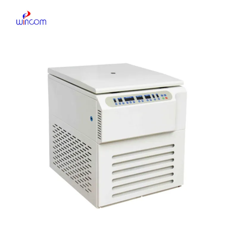
We’ve used this centrifuge for several months now, and it has performed consistently well. The speed control and balance are excellent.
To protect the privacy of our buyers, only public service email domains like Gmail, Yahoo, and MSN will be displayed. Additionally, only a limited portion of the inquiry content will be shown.
We’re looking for a reliable centrifuge for clinical testing. Can you share the technical specific...
I’m looking to purchase several microscopes for a research lab. Please let me know the price list ...
E-mail: [email protected]
Tel: +86-731-84176622
+86-731-84136655
Address: Rm.1507,Xinsancheng Plaza. No.58, Renmin Road(E),Changsha,Hunan,China

 af
af
 es
es
 ar
ar
 tr
tr
 sw
sw
 pt
pt
 th
th
 ur
ur
 bn
bn
 ne
ne
 vi
vi
 km
km
 lo
lo
 de
de
 ru
ru
 fi
fi
 nl
nl
 fa
fa
 fr
fr
 ko
ko