In the hospital and laboratory environments, mri anesthesia machine provides a complete anesthesia management solution by integrating gas delivery, ventilation support, and continuous physiological monitoring. The equipment measures oxygen saturation, airway pressures, and respiratory parameters, thus enabling anesthesiologists to react promptly to the demands of the patients. A precise control of anesthetic gases is guaranteed by integrated safety alarms and adjustable flow systems. mri anesthesia machine finds its utility in operating rooms, emergency care units, and laboratory-based clinical studies. Its dependable performance not only secures patient stability but also increases the efficiency of the procedure and facilitates uniform monitoring across various hospital settings.

mri anesthesia machine is being practiced in emergency departments as a means of fast anesthesia induction during urgent surgical interventions. Trauma cases usually have unstable patients, which makes precise management of ventilation and anesthetic delivery very important. The apparatus gives the medical personnel the opportunity to rapidly adapt gas flow and airway pressure to the patient's reaction. The introduction of the device is facilitating the management of the airway securely during the period of emergency intubation and immediate operative care. By its dependable function in emergencies, mri anesthesia machine not only helps but also hospitals to provide anesthesia that is effective during emergencies that are critically timed.
As a result of developments in laboratory and research environments, the future of mri anesthesia machine might consist of more precise experimental anesthesia protocols. Control systems that are more advanced could enable the researchers to keep the very stable environments for a long-term study. The data recording and analysis could become very complex, thus supporting the research accuracy. The above-mentioned advances would demonstrate the increasing importance of mri anesthesia machine not only in the clinical surgery but also in medical science and experimental research areas by making it more relevant.
mri anesthesia machine care is very much facilitated through proper user training. Staff in the medical sector should be well acquainted with the right procedures of startup, shutdown, and basic inspection. Wrong handling may cause extra wear and tear or may lead to performance problems. Hospitals and labs conduct training programs that further promote good daily practices. Maintenance requirements are lower if the users stick to the recommended handling methods, and the life of the equipment is longer.
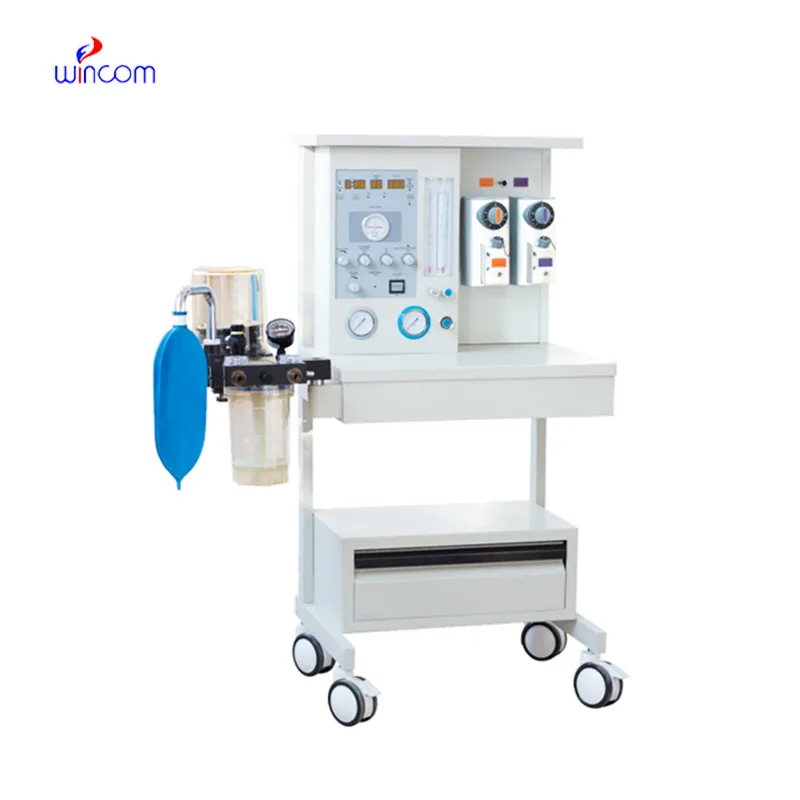
mri anesthesia machine is a crucial medical device in the operating room that supplies anesthesia controlled for patients during surgical operations. It unites accurate gas flow regulation with patient respiration and monitoring plus oxygen levels. In a hospital setting, this equipment guarantees patient safety while keeping the necessary sedation. By connecting ventilation support, vaporizer controls, and monitoring systems, mri anesthesia machine makes it possible for anesthesiologists to practice patient care easily and gives trustworthy performance in all surgical, emergency, and laboratory environments where precision in anesthesia delivery is of utmost importance.
Q: Can the Anesthesia Machine be utilized in places apart from the operating rooms? A: It can also be applied in emergency rooms, ICUs, and procedure suites. Q: What is the significance of ventilation in the anesthesia process? A: The patient’s oxygen exchange is kept at a good level during anesthesia by ventilation. Q: Are the machine’s components disposable? A: To keep the hygiene level high, it is common that breathing circuits and masks are for single use only. Q: In what ways does the machine contribute to patient safety? A: Active monitoring and alerts are there to spot the unfit conditions earlier than anyone else. Q: Is the calibration of the device done frequently? A: Calibration ensures not only the accuracy of gas delivery but also the quality of monitoring performance.
The delivery bed is well-designed and reliable. Our staff finds it simple to operate, and patients feel comfortable using it.
The water bath performs consistently and maintains a stable temperature even during long experiments. It’s reliable and easy to operate.
To protect the privacy of our buyers, only public service email domains like Gmail, Yahoo, and MSN will be displayed. Additionally, only a limited portion of the inquiry content will be shown.
Hello, I’m interested in your centrifuge models for laboratory use. Could you please send me more ...
I’m looking to purchase several microscopes for a research lab. Please let me know the price list ...
E-mail: [email protected]
Tel: +86-731-84176622
+86-731-84136655
Address: Rm.1507,Xinsancheng Plaza. No.58, Renmin Road(E),Changsha,Hunan,China

 af
af
 es
es
 ar
ar
 tr
tr
 sw
sw
 pt
pt
 th
th
 ur
ur
 bn
bn
 ne
ne
 vi
vi
 km
km
 lo
lo
 de
de
 ru
ru
 fi
fi
 nl
nl
 fa
fa
 fr
fr
 ko
ko