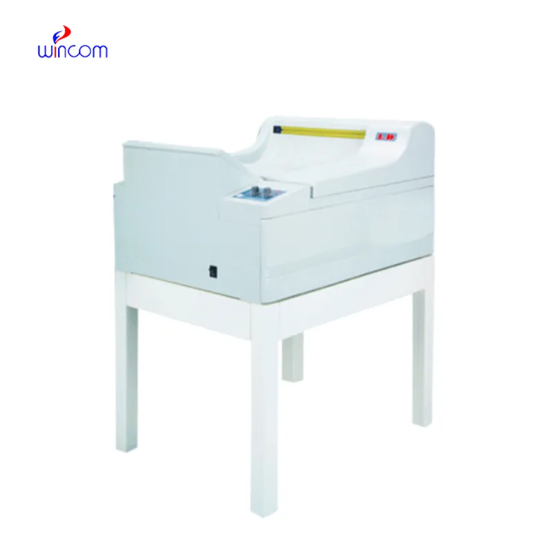
The tsa x ray scanner comes with advanced imaging sensors that ensure uniformity of images. The system also contains automatic exposure levels that ensure high images with reduced patient exposure. The tsa x ray scanner system can be adapted to suit the various functions it may be applied in. These functions include overall radiography, orthopedic images, and dental images.

The tsa x ray scanner is commonly used in medical imaging to examine skeletal trauma, lung disease, and dental anatomy. The tsa x ray scanner assists physicians in diagnosis of fractures, infection, and degenerative disease. The tsa x ray scanner is also used in orthopedic surgery intraoperatively. In emergency medicine, it provides rapid diagnostic information that allows clinicians to assess trauma and internal injury rapidly.
Future versions of the tsa x ray scanner will combine energy-efficient technology with high-resolution imaging. Predictive analytics integration will enable early disease detection and personalized screening. Global telemedicine networks will also be enabled by the tsa x ray scanner, extending access to diagnosis in underserved populations.
Maintenance of the tsa x ray scanner requires close attention to mechanical, electrical, and imaging parts. Regular visual examination catches wear or damage early. The tsa x ray scanner must be cleaned using non-abrasive substances, and filters or protective covers periodically replaced. Preventive maintenance minimizes downtime and provides reliable diagnostic results.
The tsa x ray scanner represents an important diagnostic tool that functions by using controlled X-ray radiation to create images of the bones, organs, and internal structures of the body. The equipment assists healthcare providers in diagnosing ailments such as fractures and infections with high accuracy. The tsa x ray scanner equipment is usually found in hospitals and dental clinics as it provides efficient imagery services that aid in comprehensive diagnoses. The equipment's efficiency makes it an important aspect of modern medical facilities.
Q: What are the main components of an x-ray machine? A: The main components include the x-ray tube, control panel, collimator, image receptor, and protective housing, all working together to produce diagnostic images. Q: How should an x-ray machine be maintained? A: Regular inspection, calibration, and cleaning are essential to keep the x-ray machine operating accurately and safely over time. Q: What industries use x-ray machines besides healthcare? A: X-ray machines are also used in security screening, industrial testing, and materials inspection to identify defects or hidden items. Q: Why is calibration important for an x-ray machine? A: Calibration ensures that the machine delivers accurate radiation doses and consistent image quality, which is crucial for reliable diagnostics. Q: How long does an x-ray machine typically last? A: With proper maintenance, an x-ray machine can remain operational for over a decade, depending on usage frequency and environmental conditions.
The hospital bed is well-designed and very practical. Patients find it comfortable, and nurses appreciate how simple it is to operate.
The centrifuge operates quietly and efficiently. It’s compact but surprisingly powerful, making it perfect for daily lab use.
To protect the privacy of our buyers, only public service email domains like Gmail, Yahoo, and MSN will be displayed. Additionally, only a limited portion of the inquiry content will be shown.
We’re interested in your delivery bed for our maternity department. Please send detailed specifica...
Hello, I’m interested in your water bath for laboratory applications. Can you confirm the temperat...
E-mail: [email protected]
Tel: +86-731-84176622
+86-731-84136655
Address: Rm.1507,Xinsancheng Plaza. No.58, Renmin Road(E),Changsha,Hunan,China

 af
af
 es
es
 ar
ar
 tr
tr
 sw
sw
 pt
pt
 th
th
 ur
ur
 bn
bn
 ne
ne
 vi
vi
 km
km
 lo
lo
 de
de
 ru
ru
 fi
fi
 nl
nl
 fa
fa
 fr
fr
 ko
ko