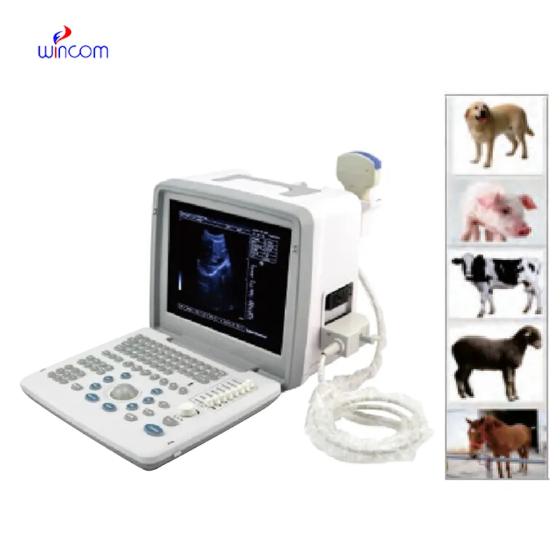
The ultrasound picture scanner, being designed for flexibility, brings the user a variety of imaging modes such as B-mode, M-mode, and color Doppler to use. Because of its small size, it can be easily moved from one place to another and that is why it can be used for examining patients in bed. The ultrasound picture scanner provides good imaging quality, which can be depended upon in cases of routine diagnostics, fieldwork, and emergency medical interventions.

The ultrasound picture scanner is the workhorse in oncology because it assists in the accurate locating of tumors and keeping an eye on the treatment progress. It helps in making the right calls concerning benign versus malignant lesions in breast and thyroid cases. The ultrasound picture scanner is also there in support of interventional procedures such as guided aspirations and injections.

The coming years will see the evolution of the ultrasound picture scanner into an independent and adaptable imaging solution. The increased level of automation will eliminate the need for human input. The ultrasound picture scanner may include predictive model components that will help healthcare providers to identify probable risks to an individual's health.

In order to extend the service life of the ultrasound picture scanner, it is recommended that users refrain from applying much force during the process of connecting/disconnecting probes. Power cables should always remain dry. The ultrasound picture scanner needs diagnostic tests to ensure that it produces quality images.
Built for performance and accuracy, the ultrasound picture scanner is an imaging diagnosis platform. It gives real-time images of tissues, organs, and vascular systems, enabling increased detection of pathology. Space-efficient and compact, the ultrasound picture scanner is ideal for hospitals, clinics, and ambulatory healthcare facilities. Its accuracy imaging enables physicians to offer timely and informed medical care.
Q: What makes the ultrasound scannert effective for diagnostic imaging? A: Its high-frequency sound wave technology allows accurate visualization of internal body structures in real time. Q: How portable is the ultrasound scannert? A: The device features a compact and lightweight design, allowing easy movement between clinical departments. Q: What types of probes are compatible with the ultrasound scannert? A: It supports multiple probe types, including linear, convex, and phased array probes for varied diagnostic needs. Q: Does the ultrasound scannert require special training to operate? A: Basic technical training is recommended to maximize its imaging performance and functionality. Q: How long can the ultrasound scannert operate continuously? A: It is designed for extended use with efficient cooling systems and stable power performance.
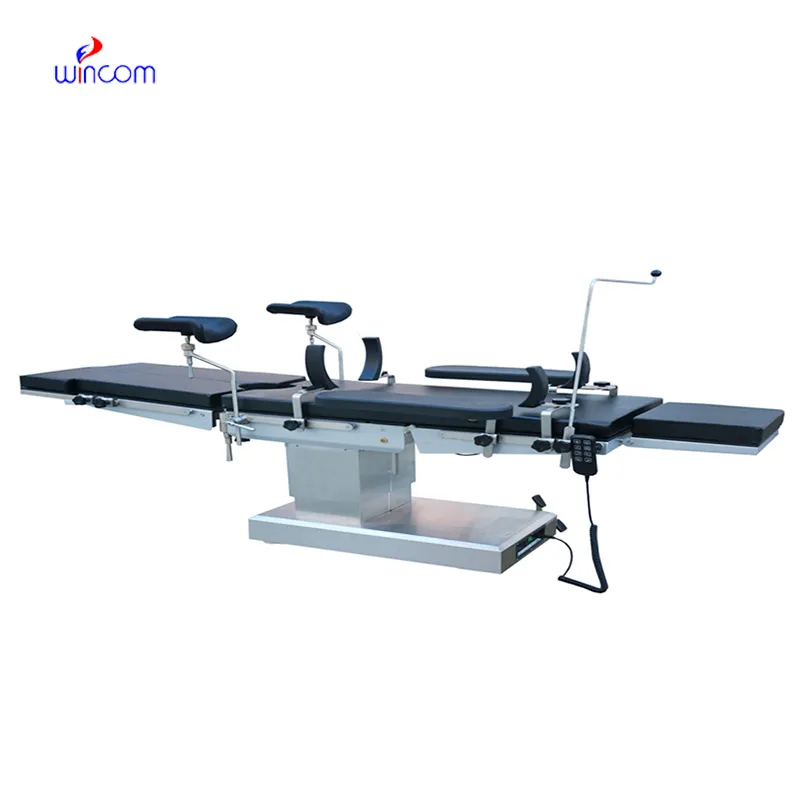
The hospital bed is well-designed and very practical. Patients find it comfortable, and nurses appreciate how simple it is to operate.
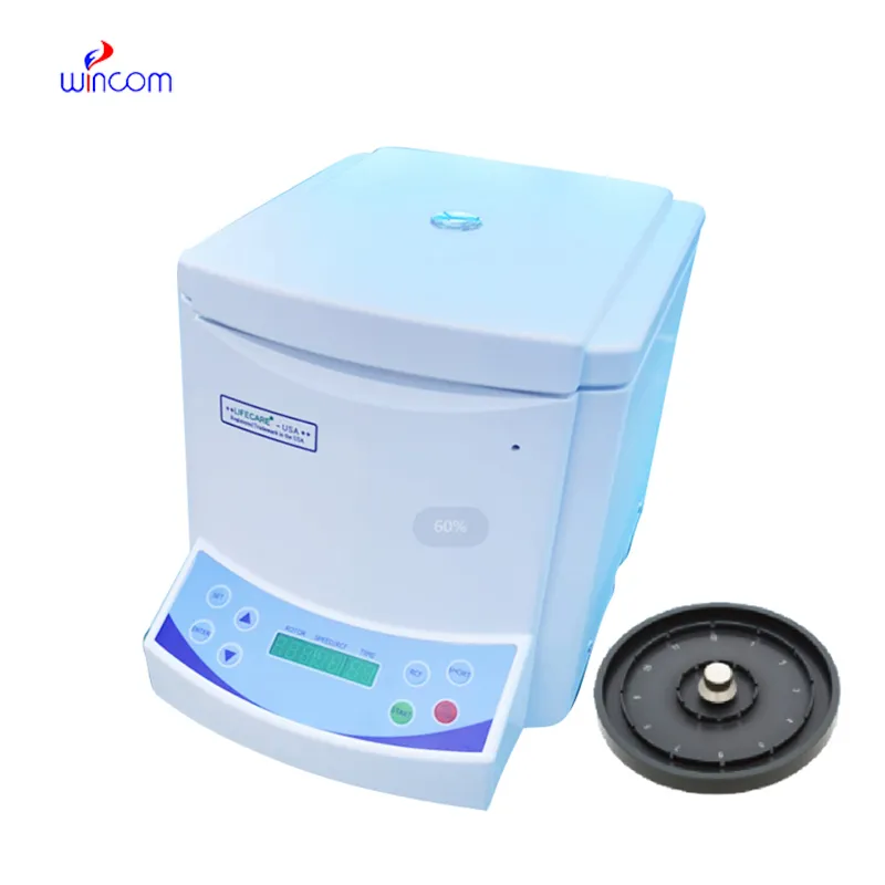
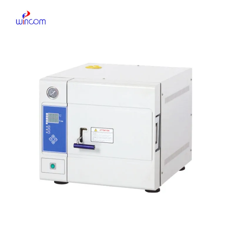
The centrifuge operates quietly and efficiently. It’s compact but surprisingly powerful, making it perfect for daily lab use.
To protect the privacy of our buyers, only public service email domains like Gmail, Yahoo, and MSN will be displayed. Additionally, only a limited portion of the inquiry content will be shown.
Hello, I’m interested in your centrifuge models for laboratory use. Could you please send me more ...
Hello, I’m interested in your water bath for laboratory applications. Can you confirm the temperat...
E-mail: [email protected]
Tel: +86-731-84176622
+86-731-84136655
Address: Rm.1507,Xinsancheng Plaza. No.58, Renmin Road(E),Changsha,Hunan,China

 af
af
 es
es
 ar
ar
 tr
tr
 sw
sw
 pt
pt
 th
th
 ur
ur
 bn
bn
 ne
ne
 vi
vi
 km
km
 lo
lo
 de
de
 ru
ru
 fi
fi
 nl
nl
 fa
fa
 fr
fr
 ko
ko